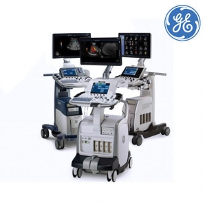
GE Healthcare LOGIQ Vision Series Ultrasound System, Model Name/Number: Logiq V3 And Logiq V5
OUT OF STOCK
ISO Certified
ISO 9001:2001
 Precult Delivery
 Contactless Delivery
Get Quotation
Overview:
- Your commitment to patients and the utility of ultrasound imaging go hand in hand in building a healthier world.
- The new LOGIQ* Vision Series, including the LOGIQ V5 and the LOGIQ V3 consoles, makes advanced ultrasound technology simple, affordable, and accessible for general imaging, shared service and OB/GYN healthcare providers.
- The systems support you with automated calculations, onboard clinical assistance, and time-saving workflow that helps enhance your confidence.
MLA4 Technology increases frame rate by 4X1 by processing each echo along four simultaneous receiver beams to study moving organs and blood flow data
Real-Time Speckle Reduction Imaging (SRI-HD) helps reduce noise while enhancing true tissue architecture
CrossXBeam* imaging enhances tissue interfaces and border differentiation with compound imaging technology
Coded Harmonic Imaging helps improve near-field resolution in small-parts imaging
B-Steer helps view deep vessels and structures that are angled (ex. tendons insertion)
Perform advanced clinical evaluations that help you diagnose confidently across a wide range of applications:
Advanced imaging modes like Color°°, Pulse Wave and Power Doppler Imaging (PDI)°° help measure blood flow within a specific area to assist with diagnosis and monitoring
Anatomical M-Mode° supports anatomical measurements in non-standard planes
LOGIQ View° allows real-time imaging of long anatomical structures too large to be seen in a single image
Virtual Convex provides expanded field of view when using linear and sector probes
Easy 3D° Technology helps acquire freehand 3D dataset to visualize volumetric anatomies
Experience Simplicity and Speed throughout your daily routine with the LOGIQ V5 and LOGIQ V3:
Switch Probe button to quickly switch between the two active probes
User Preset button to quickly create or overwrite user presets
Auto IMT* provides automatic edge detection for intima-media thickness (IMT)
SonoBiometry is a workflow tool that automatically places calipers to help you perform key fetal biometry measurements quickly
TruScan™ Architecture (GE Raw Data) allows limitless flexibility to readjust a large set of imaging parameters and re-measure images any time after you have set, frozen or even archived the images